
Keratoconus: Early Detection and Management in Seoul
How Seoul patients can catch keratoconus early — and keep it from progressing
Q. What is keratoconus?
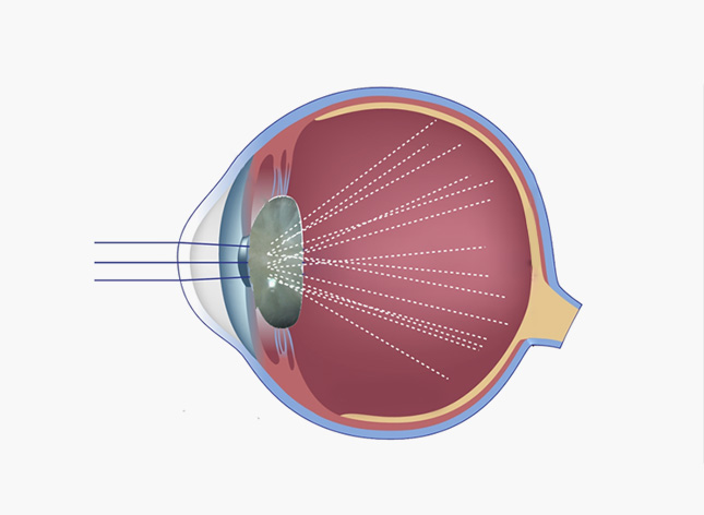
Keratoconus is a progressive eye disease in which the normally round, dome-shaped cornea gradually thins and bulges outward into an irregular cone shape. This distorts the way light enters the eye, causing blurred and distorted vision, glare, and frequent changes in glasses prescription that eyeglasses alone often cannot fully correct.
Q. Who develops keratoconus and why?
Keratoconus most often begins during the teenage years or early twenties and progresses gradually over one to two decades before stabilizing, typically in a person’s thirties or forties. It is associated with a genetic predisposition, chronic eye rubbing, allergic conjunctivitis, and certain connective tissue disorders. In Seoul, where allergic conjunctivitis and heavy near-work are common among students and office workers, unexplained or rapidly changing astigmatism should always prompt a corneal topography scan rather than simply a new glasses prescription.
Q. What are the symptoms and signs of keratoconus?
- Blurred or distorted vision that changes over time, with frequent, unexplained changes in glasses prescription
- Increased sensitivity to light and glare, especially when driving at night
- Halos or streaking around lights, making night driving increasingly difficult
- Frequent eye rubbing or itching, often related to underlying seasonal or perennial allergies
- Progressive astigmatism that soft contact lenses or glasses can no longer fully correct
Keratoconus by the numbers
▸ Classic estimates place keratoconus at roughly 1 in 2,000 people, but modern Scheimpflug corneal imaging studies suggest true prevalence may be as high as 1 in 375, largely due to improved early detection.
▸ Onset commonly occurs in the late teens to mid-twenties, with a modest male predominance reported across East Asian populations.
▸ Korea has one of the highest myopia rates in the world among young adults, and overlapping refractive complaints can mask early keratoconus if corneal topography is never performed.
▸ Chronic eye rubbing — common among patients with allergic conjunctivitis and atopic dermatitis, both frequently seen across Yeongdeungpo, Guro, Gasan, and Gwanak — remains one of the strongest modifiable risk factors for progression.
Q. How is keratoconus treated?
Corneal cross-linking is the standard first-line treatment once topography confirms progression. Riboflavin drops combined with controlled UV-A light exposure strengthen the bonds between corneal collagen fibers, halting further steepening in the majority of treated eyes when performed early.
Rigid gas permeable, hybrid, or scleral contact lenses vault over the irregular corneal surface to restore clear vision once glasses are no longer sufficient. In advanced cases with significant scarring, a corneal transplant such as DALK or penetrating keratoplasty may become necessary.
Q. Can keratoconus be managed without surgery in its early stages?
Yes. Many patients diagnosed early through corneal topography can be managed for years with specialty contact lenses, regular monitoring scans, and active allergy control to reduce eye rubbing. UV protection with sunglasses is also recommended, as ultraviolet exposure may contribute to corneal weakening over time. Regular follow-up imaging, often every six to twelve months, allows an ophthalmologist to catch any renewed progression early enough for cross-linking to remain an option.

⚠ When keratoconus risk should prompt a same-week eye exam
- Vigorous eye rubbing, especially at night or in association with allergies, is strongly linked to keratoconus onset and progression
- A family history of keratoconus warrants earlier corneal topography screening, ideally before any refractive surgery is considered
- Rapid, repeated changes in glasses prescription — especially increasing astigmatism — in teenagers or young adults should prompt topography testing
- Undiagnosed keratoconus is a leading cause of poor outcomes after refractive surgery, making corneal imaging before any LASIK, PRK, or LASEK procedure essential
Early Stage
Mild irregular astigmatism, often still correctable with glasses or soft contact lenses. Topography shows subtle inferior corneal steepening.
Moderate Stage
Progressive thinning and steepening. Glasses are no longer sufficient; rigid gas permeable or scleral lenses are typically required, and cross-linking is strongly considered.
Advanced Stage
Significant scarring and thinning with possible contact lens intolerance and risk of acute hydrops. Corneal transplant may be considered.
“In my clinic in Yeongdeungpo, I regularly see young patients referred after their glasses prescription changed several times within a single year. Corneal topography takes only a few minutes, and catching keratoconus early with corneal cross-linking can preserve vision for decades. I encourage anyone with a family history of keratoconus, or unstable astigmatism, to have their corneal shape mapped rather than simply prescribing another new lens.”
— Dr. Daniel Baik, Nunehim Eye Center

❓ Frequently Asked Questions
Q. Is keratoconus hereditary?
Keratoconus has a clear genetic component. Having a first-degree relative with the condition raises an individual’s risk substantially compared with the general population, so family members of diagnosed patients are advised to begin corneal topography screening in their teenage years rather than waiting for symptoms to appear.
Q. Does keratoconus always require surgery?
No. Many patients with mild to moderate keratoconus achieve clear, comfortable vision with glasses or specialty contact lenses such as rigid gas permeable or scleral lenses. Corneal cross-linking is recommended once topography confirms the condition is progressing, and corneal transplant is reserved for advanced cases with significant scarring or thinning.
Q. Can LASIK be performed if I have keratoconus?
Standard LASIK is not recommended for eyes with keratoconus because removing corneal tissue from an already thinned and weakened cornea can accelerate its bulging. Surface treatments such as TransPRK or 2-Day LASEK carry a lower but still meaningful risk, which is why thorough corneal topography screening before any refractive surgery in Seoul is essential.
Q. How is keratoconus diagnosed?
Diagnosis relies on corneal topography or tomography, imaging technology that maps the front and back curvature of the cornea in detail. This allows an ophthalmologist to detect the asymmetric steepening and thinning characteristic of keratoconus, often years before it would be noticeable on a standard eye exam or vision test.
Q. What is corneal cross-linking and how effective is it?
Corneal cross-linking uses riboflavin eye drops activated by ultraviolet-A light to create additional bonds between the collagen fibers that make up the cornea, strengthening the tissue. Performed at an early stage, it halts further steepening in the large majority of treated eyes and is considered the standard first-line treatment for documented progression.
Q. Can children get keratoconus?
Yes, and pediatric keratoconus tends to progress faster and more aggressively than the adult-onset form of the disease. Children who rub their eyes frequently due to allergies, or who have a family history of keratoconus, should be screened earlier with corneal topography and monitored more closely than adult patients.
Q. Where can I get a corneal topography screening in Seoul?
Nunehim Eye Center in Yeongdeungpo-gu offers corneal topography and keratoconus evaluation under Dr. Daniel Baik, serving patients from Yeongdeungpo, Guro, Gasan, and Gwanak in both Korean and English consultations.

Book a Consultation
nunehim.com — Yeongdeungpo · Guro · Gasan · Gwanak
📍 Visit Nunehim Eye Center — Guro, Seoul
Convenient access from Guro, Gasan, Gwanak & Dongjak. Near Guro Digital Complex Station (Line 2).
Address: 551 Siheung-daero, Guro-gu, Seoul — nunehim.com
Hours: Mon–Fri 09:00–18:30 (lunch 13–14), Sat 09:00–14:00
Languages: Korean & English


Leave a comment