
Macular Pucker: Symptoms, Diagnosis and Treatment
Macular pucker care in Yeongdeungpo, Guro & Gasan, Seoul
Q. What is an epiretinal membrane?
An epiretinal membrane (ERM), also called macular pucker, is a thin layer of scar-like tissue that forms on the surface of the macula and distorts central vision. Most cases in patients from Yeongdeungpo, Guro, and Gasan are mild and only need monitoring, but surgery — a vitrectomy with membrane peeling — is recommended when distortion or vision loss begins to affect daily life.
Q. What causes an epiretinal membrane to form?
An epiretinal membrane most often develops after the vitreous gel inside the eye separates from the retina with age (posterior vitreous detachment), leaving behind cells that proliferate into a thin fibrous layer on the macula. It can also follow retinal tears, uveitis, diabetic retinopathy, retinal vein occlusion, or previous eye surgery. Idiopathic ERM — with no identifiable underlying cause — is most common in patients over 50 to 60.
▸ Population-based studies in Asian populations estimate idiopathic epiretinal membrane affects roughly 2–6% of adults over 50, rising to more than 10% in those over 70.
▸ Retina clinics in Korea report a growing number of ERM diagnoses as the country’s population aged 65 and older continues to expand rapidly.
▸ Only a minority of diagnosed ERM cases — roughly 10–20% — progress enough to require surgery within five years of diagnosis.
▸ Bilateral ERM occurs in a meaningful subset of patients, around 20–30%, so both eyes should be monitored even when only one is symptomatic.
Q. What are the symptoms of an epiretinal membrane?
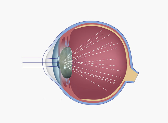
The hallmark symptom is metamorphopsia — straight lines, door frames, or text appearing wavy or bent. Patients also commonly notice blurred or reduced central vision and difficulty reading fine print, and in some cases mild monocular double vision.
Symptoms are usually mild and progress slowly over months to years, which is why regular eye exams matter for patients across Guro and Gasan even when vision still feels ‘good enough.’
Q. How is an epiretinal membrane diagnosed?
A dilated fundus exam often reveals a glistening membrane and fine wrinkling on the retinal surface. Optical coherence tomography (OCT) is the gold-standard test, confirming the diagnosis and precisely measuring membrane thickness and associated macular swelling.
An Amsler grid test can also help patients monitor visual distortion at home between clinic visits, flagging any worsening that should prompt an earlier follow-up.

Q. When does an epiretinal membrane need surgery?
- Progressive worsening of visual acuity
- Significant metamorphopsia affecting reading, driving, or work
- OCT evidence of increasing macular thickening or traction
- Symptoms affecting quality of life or binocular vision
- High visual demands from occupation or hobbies
Stages of Epiretinal Membrane
Mild
Thin membrane, minimal symptoms: annual OCT monitoring
Moderate
Visible distortion, mild vision loss: monitor every 6 months
Advanced
Significant metamorphopsia or vision loss: vitrectomy recommended
A Note from Dr. Daniel Baik
Many of my patients in Yeongdeungpo and Gasan first notice an epiretinal membrane when straight lines start to look wavy while reading. I always reassure them that most cases can simply be monitored — surgery is reserved for eyes where distortion truly affects daily life, and modern vitrectomy techniques offer good visual recovery. — Dr. Daniel Baik, board-certified ophthalmologist

❓ Frequently Asked Questions
Q. Is an epiretinal membrane the same as a macular hole?
No. An epiretinal membrane is a fibrous layer sitting on top of the macula, while a macular hole is a break in the retinal tissue itself. They can sometimes occur together, but they are distinct conditions with different management.
Q. Will an epiretinal membrane go away on its own?
Rarely. Most epiretinal membranes remain stable or progress very slowly, and they do not resolve without surgical removal. Regular monitoring is used to track whether treatment eventually becomes necessary.
Q. What does vitrectomy with membrane peeling involve?
It is typically an outpatient or short-stay surgery in which the vitreous gel is removed and the membrane is gently peeled from the macular surface, often together with the internal limiting membrane (ILM) to reduce the chance of recurrence.
Q. How long is recovery after ERM surgery?
Vision typically improves gradually over several weeks to months as retinal swelling resolves. Some residual distortion can persist, especially if the membrane was present for a long time before surgery.
Q. Can an epiretinal membrane come back after surgery?
Recurrence is uncommon, particularly when the internal limiting membrane is peeled at the time of surgery, though your surgeon will continue periodic OCT monitoring afterward.
Q. Does an epiretinal membrane affect both eyes?
It can. A meaningful subset of patients, roughly 20–30%, develop ERM in both eyes, so ophthalmologists typically monitor the fellow eye even if it is not yet symptomatic.
Q. Where can I get an OCT evaluation for epiretinal membrane near Guro?
Nunehim Eye Center in Guro-gu, Seoul offers OCT imaging and comprehensive retinal evaluation for patients from Yeongdeungpo, Guro, Gasan, and Gwanak, with both Korean and English consultations available.

Book a Retina Consultation
📍 Visit Nunehim Eye Center — Guro, Seoul
Convenient access from Guro, Gasan, Gwanak & Dongjak. Near Guro Digital Complex Station (Line 2).
Address
551 Siheung-daero, Guro-gu, Seoul / nunehim.com
Hours
Mon–Fri 09:00–18:30 (lunch 13–14), Sat 09:00–14:00
Languages
Korean & English


Leave a comment