Red, Swollen Inner Corner of Your Eye? Understanding Carunculitis
A board-certified ophthalmologist’s guide from Nunehim Eye Center, Guro, Seoul
Q. What is carunculitis, in one sentence?
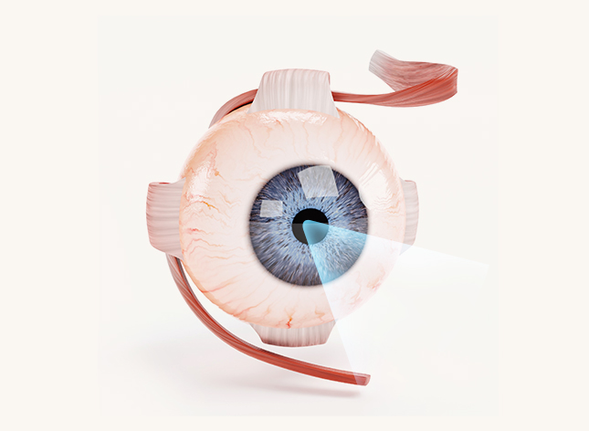
Carunculitis is inflammation of the lacrimal caruncle — the small pink tissue at the inner corner of the eye, near the nose — and while it can feel similar to a stye, punctitis, or canaliculitis, it is a distinct condition that needs a different diagnostic approach.
Q. Where exactly is the lacrimal caruncle?
If you look closely at the inner corner of your eye, nearest your nose, you’ll see a small piece of pink tissue — this is the lacrimal caruncle. It looks like a simple bit of conjunctiva, but it is actually a fairly complex structure containing conjunctival tissue, skin-like tissue, sebaceous glands, sweat glands, hair follicles, and accessory lacrimal gland tissue all in one small area. Because so many different tissue types are packed into this spot, almost any type of lesion that can occur in skin, conjunctiva, or glandular tissue can also occur in the caruncle — which is part of why caruncular lesions are so varied.
Q. Is carunculitis the same as punctitis or canaliculitis?
No. These are three different structures that happen to sit very close together. The caruncle is the pink tissue described above. The punctum is the small drainage opening on the inner edge of the upper and lower eyelids where tears exit the eye. The canaliculus is the tear duct that carries tears from the punctum toward the nose. Inflammation of any of these three — carunculitis, punctitis, or canaliculitis — can feel like general ‘pain in the inner corner of the eye’ to a patient, which is exactly why an ophthalmologist needs to identify the precise structure involved before treating it.
Q. What are the symptoms of carunculitis?
- Redness at the inner corner of the eye
- Visible swelling of the caruncle itself
- Stinging or tenderness when touched or blinking
- A foreign-body sensation
- Increased tearing
- Discharge or crusting
- Rarely, a pus-filled abscess
The key distinguishing feature is location: if the small pink structure at the inner, nasal corner of the eye is swollen — rather than the eyelid margin itself — a caruncular lesion should be considered.
What the Medical Literature Tells Us
- ▸ Carunculitis is not a condition with large standardized studies behind it — the available literature is mostly case reports and histopathology studies of inflammatory lesions, infected cysts, and abscesses of the caruncle
- ▸ A documented case of an Actinomyces caruncular abscess was successfully treated with incision, drainage, and antibiotics
- ▸ Caruncular swelling can also appear as one of the active inflammatory signs of thyroid eye disease
- ▸ Histopathology studies have found a notable mismatch between clinical impression and final biopsy diagnosis for caruncular lesions — meaning a lesion that looks like simple inflammation is not always confirmed as such once examined under a microscope
Q. What causes carunculitis?
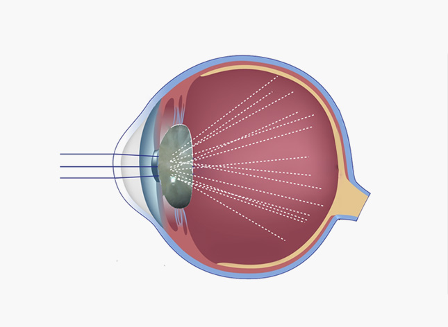
Because the caruncle contains sebaceous glands and hair follicles, localized inflammation or infection can develop there, occasionally progressing to an abscess. Blocked glands or ducts can also lead to retention cysts or epidermoid cysts, which become red and painful if they become infected or inflamed.
Since the caruncle sits directly against the conjunctiva, nearby conjunctival inflammation or ongoing surface irritation can make the area look swollen as well. Rarely, caruncular swelling is linked to a systemic condition such as thyroid eye disease or an immune-related process — which is why recurring or bilateral swelling shouldn’t automatically be assumed to be a simple local infection.
Q. How is carunculitis different from a stye?
A stye (hordeolum) typically develops along the eyelid margin, involving the meibomian or Zeis glands, and appears as a localized bump. Carunculitis instead involves swelling of the caruncle itself — the pink tissue in the inner corner — and requires a slit-lamp exam to properly distinguish it from a cyst, tumor, or canaliculitis rather than simply treating it as a stye.
Q. When should you see an ophthalmologist?
- The lesion persists for more than 2 weeks
- It is gradually growing in size
- You can feel a firm or hard lump
- The surface bleeds easily
- It is darkening or changing color
- The same spot keeps swelling repeatedly
- It isn’t improving with antibiotic treatment
- Pus or discharge continues
Because clinical appearance and final biopsy-confirmed diagnosis often don’t match for caruncular lesions, a lesion that lingers should be evaluated rather than self-treated indefinitely.
Benign Lesions
- Nevus (pigmented mole)
- Papilloma
- Pyogenic granuloma
- Retention or epidermoid cysts
Rare Malignant Lesions
- Basal cell carcinoma
- Sebaceous carcinoma
- Melanoma
Work-up
- Slit-lamp examination
- Punctum & canaliculus check
- Biopsy if persistent or atypical
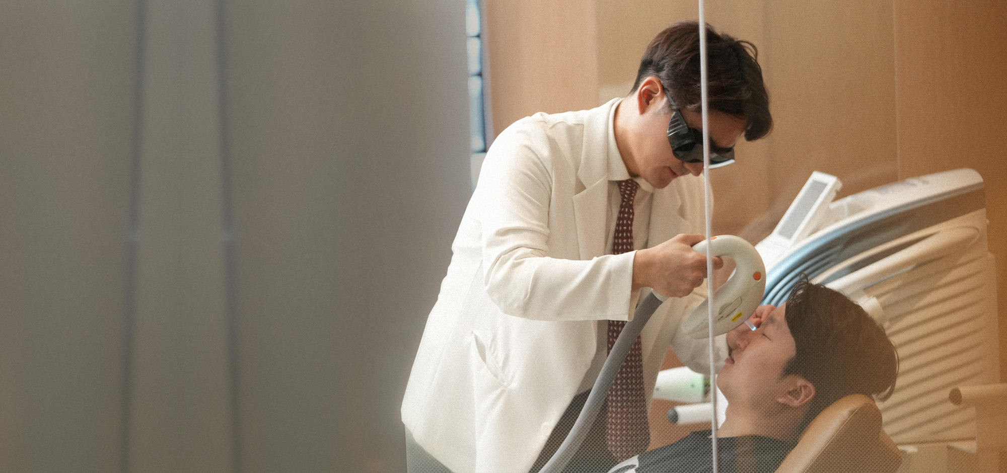
“In my clinic in Guro, I sometimes see patients who assumed they had a stye and self-treated for weeks before coming in, when the actual problem was a cyst or an inflammation specific to the caruncle itself. Because this small structure contains so many different tissue types, an accurate slit-lamp exam matters far more than guessing from a mirror.” — Dr. Daniel Baik, board-certified ophthalmologist
Q. How is carunculitis treated?
Treatment depends on the underlying cause rather than a single default approach. Mild irritation or inflammation is often managed conservatively with artificial tears and avoiding touching or rubbing the area. If a bacterial infection is suspected, topical antibiotic drops or ointment may be prescribed based on the exam findings. An abscess or infected cyst may not respond to drops alone and can require incision and drainage or removal of the lesion — as in the reported Actinomyces abscess case, which was treated with surgical drainage plus antibiotics. If a mass persists or the diagnosis is uncertain, excisional biopsy may be needed to rule out papilloma, cyst, nevus, or a tumor.
Q. Does a warm compress help?
Not universally. A warm compress can help if the swelling is related to blocked sebaceous glands or accompanying blepharitis, but it will not resolve a cyst, canaliculitis, abscess, or tumor-like lesion. If there’s no improvement after a few days of warm compresses, it’s time to get evaluated rather than continuing home care.
What Not to Do
- Don’t squeeze or press the area with your fingers — the punctum and canaliculus sit right next to the caruncle
- Don’t use leftover steroid drops without guidance — they can worsen an underlying infection
- Don’t reuse old antibiotic drops left over from a previous stye
- Stop wearing contact lenses while the area is actively inflamed, and switch to glasses in the meantime
❓ Frequently Asked Questions
Q. Does any redness at the inner corner of the eye mean carunculitis?
Not necessarily. The caruncle can develop a wide range of lesions besides inflammation, including cysts, nevi, papillomas, and pyogenic granulomas. A lesion that persists or grows should be examined with a slit lamp rather than assumed to be simple inflammation.
Q. Is carunculitis the same as punctitis?
No. The caruncle is the pink tissue at the inner corner of the eye, while the punctum is the tear-drainage opening on the eyelid. They sit close together and are easy to confuse, but they are different structures.
Q. How can I tell it apart from a stye?
A stye usually develops along the eyelid margin. Carunculitis instead involves swelling of the pink caruncle tissue itself, in the corner nearest the nose.
Q. Will it go away on its own?
Mild irritation can resolve on its own. But a lesion that persists or keeps recurring in the same spot may not be simple inflammation, and should be evaluated.
Q. Is carunculitis contagious?
Localized inflammation of the caruncle itself isn’t considered a contagious condition. However, if it occurs alongside viral or bacterial conjunctivitis, that underlying conjunctivitis may be contagious, so good hand hygiene is still important.
Q. Is an antibiotic eye drop enough to treat it?
Not always. Antibiotics may help if there’s a bacterial infection, but cysts, abscesses, canaliculitis, or tumor-like lesions need different treatment. Identifying the actual cause first is essential.
Q. The swelling keeps getting bigger — should I be concerned?
Simple inflammation typically improves over time. A lesion that keeps growing over several weeks should be evaluated to rule out other types of lesions.
Book a Consultation
If the inner corner of your eye has been red, swollen, or painful for more than a few days, Nunehim Eye Center offers Korean & English consultations in Guro, Seoul.
Or visit nunehim.com
📍 Visit Nunehim Eye Center — Guro, Seoul
Convenient access from Guro, Gasan, Gwanak & Dongjak. Near Guro Digital Complex Station (Line 2).
Address: 551 Siheung-daero, Guro-gu, Seoul / nunehim.com
Hours: Mon–Fri 09:00–18:30 (lunch 13:00–14:00), Sat 09:00–14:00
Languages: Korean & English


Leave a comment